OLFACTORY GROOVE MENINGIOMAS
Consideration of this "classical" location for a meningioma, extensively studied by H.Cushing, brings together effectively all the principles of a meningioma's removal. Attachment is on the cribriform plate, affecting olfactory bulbi and tracts, in major lesions extending towards crista galli and planum sphenoidale.
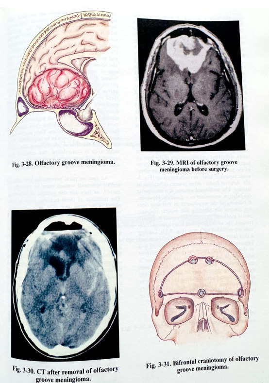
The tumour tends to occupy the inter-hemispherical fissure, indenting the frontal lobes from the base and medially, and being at least several centimetres in diameter. Since, even with substantial growth and significant frontal lobes compression and displacement, patients remain virtually asymptomatic. Often they present only with anosmia and mild personality changes that are easily neglected. Its blood supply is provided from the meningeal, ethmoidal and ophthalmic arteries. Part of the tumour can invade the ethmoidal paranasal cells. Tumour growth displaces the A2 arterial segments posteriorly and superiorly, and in big tumours they are close to and even embedded in the tumour surface. Although involved, these arterial segments can practically always be separated by dissection. This is not the case with the frontopolar branches, which often must be sacrificed. Olfactory nerves are severely distended and displaced. Posteriorly, the chiasmatic cistern is also compressed and displaced (Figs. 3-28; 3-29, 3-30).
As in all meningiomas, preoperative investigations begin with detailed contrast and non-contrast CT scans, performed also as direct coronal scans. The contour of the tumour and its relationship to the hemispheres, paranasal sinuses (ethmoid and sphenoid) and falx is delineated. The "bone" window CT images show the presence and size of a hyperostosis. MRI studies give better presentation of the position of arteries, optic nerves, chasm and the extent of perifocal oedema. Angiography can be reserved only for bigger tumours and it can be helpful for the identification of feeders, and to give additional information about major adjacent arteries and veins. Preoperative planning takes into consideration the size of the tumour, and its precise attachment before selecting the approach. Big and more anteriorly attached lesions are approached bifrontally, leaving the unilateral subfrontal or pterional way for small and posteriorly located tumours.
BIFRONTAL APPROACH
The bifrontal approach is performed in the supine position, with the upper part of the body elevated and the head slightly deflected. Skin incision follows the anterior hairline, forming a flap to be turned anteriorly towards the face, together with the epicranial layer. The bone flap extends over both frontal areas, close to the most anterior part of the superior temporal line with a height of 6 cm or more (larger in huge tumours). One or two small burr holes can be placed on both sides of the superior sagittal sinus area for safe bone undercutting (Fig. 3-31). Similar special care is taken in undercutting the bone flap close to the base midline, where the bone thickening as a crest inside requires special attention to preclude perforating the dura. As basal bone cutting lines are as close to the base as possible, the craniotomy widely opens the cavities of the frontal sinuses, and special care should be taken to isolate them from the aerated spaces below, and after that the dura to be opened. The mucosa of opened sinuses is removed and the sinuses are packed with antibiotic soaked Gelfoam. An periosteal flap can be turned over the exposed sinuses and sutured to the dura, isolating them during the next stages of surgery. The bone flap is a "free" one and immediately after its lifting, care is taken to deal with any superior sagittal sinus area bleeding. This is easily stopped with oxidised cellulose and moderate elevation of the head.
The dura is opened on both sides of the falx, 3-4 cm lateral from midline and 1- 1.5 cm from the basal craniotomy edge. Both incisions are enlarged to the midline, carefully approaching the falx and superior sagittal sinus. After safely reaching it, the medial surfaces of both frontal hemispheres are dissected from the falx and retracted with small spatulas, coagulating the bridging veins. The superior sagittal sinus and the falx are divided at their most basal point between two clips or ligatured, paying attention to the inner edge of falx where venous collectors can be found. The full mobility of the falx allows the retraction of the frontal lobes. They are lifted and slightly separated at the interhemispherical plane, until the anterior part of the tumour is seen. The arachnoid is dissected to determine the proper plane of separation of cortical vessels from the tumour.
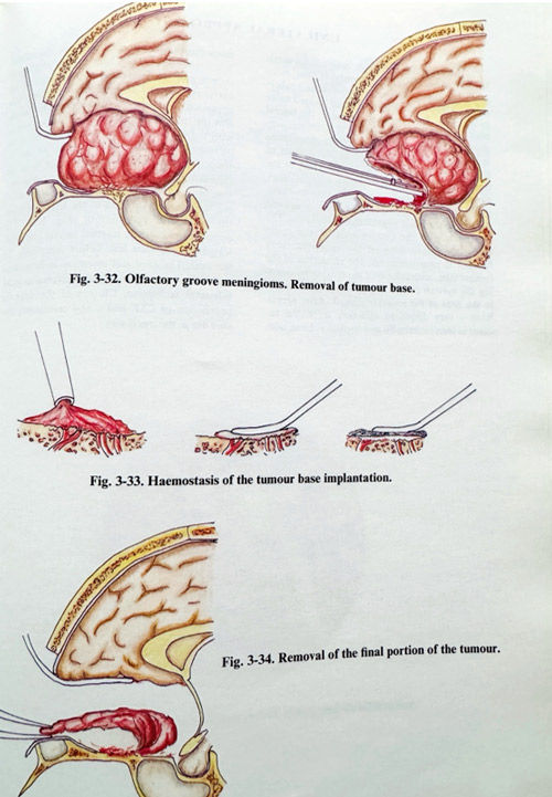
The classical advice of H.Cushing remains valid for tumour removal. After biopsies are taken, debulking and transsection of the base go together, removing bigger and bigger portions of the tumour, using ordinary suction, an ultrasonic aspirator, laser, or diathermy loop (Fig. 3-32). Arterial feeders often present difficulties in control. The traditional solution is a monopolar coagulation, connected to a dissector. However, if burning of the attachment in this way does not control the bleeding and bipolar coagulation is inefficient (it coagulates vessels inside soft tissues), the already exposed bony arterial channels are occluded with wax (Fig. 3-33). Leaving a thin layer of tumour over the attachment and hyperostosis can avoid the time and blood loss at this stage and postpone it for the moment when all the tumour mass is out, and space is much greater, the normal dural edges are seen and bleeding control can be faster. Debulking and detachment advance along the skull base in the cribriform palate area and the olfactory bulbs are rarely spared (may be only one of them at times). If both bulbs are sacrificed, both frontal lobes are retracted without limitations, but if one of them is preserved, the opposite lobe is predominantly retracted. Once the tumour has been debulked, the bleeding decreases, and the surgeon can follow the lateral border of the tumour to reach the lesser sphenoid wing and presumably the anterior clinoid area. The arachnoid is usually thick and provides protection of neural and vascular structures located behind it, but the translucent layer sometimes permits the identification of chiasm, optic nerves and supraclinoid carotids (Fig. 3-34).
Complete preservation of the cisternal arachnoid should be attempted. The posterior dome of the tumour, even in big tumours, is separated from A2 arterial segments, but the most upper part can involve (and sometimes encase) frontopolar arteries. They can be sacrificed if dissection proves to be impossible. After all parts of the tumour are removed, the attachment area is coagulated, the affected dura removed, and permanent haemostasis of feeders achieved after drilling the hyperostotic bone.
All tumour parts penetrating into ethmoid and/or sphenoid sinuses must be radically removed, as they are sources of recurrence. The resection of these portions must be completed at the same stage. The wide communication with paranasal sinuses is repaired with fascia - suturing it to a preserved part of the dura and gluing it to prevent CSF rhinorrhoea. Closure entails repairing the falx, simply tying the superior sagittal sinus ligatures one to an-other, and closing the dura along the incision.
Special care is taken to ensure water tight closure, as a CSF fistula can be a serious and more frequent complication in this type of meningioma removal. The bone flap is replaced and fixed, and epicranial layers are sutured in the usual way, leaving an epidural drain under mild suction.
UNILATERAL APPROACH
The unilateral approach, designed for smaller and more posterior tumours, is the one more often applied in planum sphenoidale and tuberculum sellae meningiomas or those located more unlaterally. The craniotomy exposes the lateral part of the frontal, pterional or anterior temporal regions after a skin incision from the midline at the anterior hairline to a point just anterior to the tragus. The flap can be left attached to the temporal muscle or be lifted as a "free" flap.
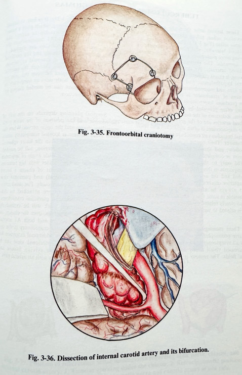
Frontoorbital craniotomy is very convenient for this purpose (Fig. 3-35). The dura is opened parallel to the basal edge of the craniotomy. The approach uses the convenience of the route along the lesser wing of the sphenoid, very familiar to neurosurgeons. The retraction of the frontal lobe, with release of the tension by opening the sylvian cistern if necessary, gives access to the area of the anterior clinoid. After identifying a very displaced olfactory tract, the tumour is seen anteriorly and medially. Even with more posteriorly attached tumours, the accessibility and safe separation of the chiasmatic cistern and the ipsilateral internal carotid are early and better, compared to the bifrontal approach. A tumour sample is taken for biopsy and the tumour is debulked in a similar way as with the bifrontal technique, but very often the olfactory tract must be sacrificed, as, due to retraction, the bulb tends to disrupt from the cribriform area. After debulking, the safe dissection of the residual part should be done following the posterior margin of the lesion, between the arachnoid and tumour (Fig. 3-36). The opposite optic nerve, internal carotid and olfactory tract are seen last, before completing the removal. The care and attention to hemostasis and radicalism of removal are essential as in the bifrontal technique. Closure of the dura for prevention of CSF leaks, and craniotomy closure are done in the usual way.