CLINOID MENINGIOMAS
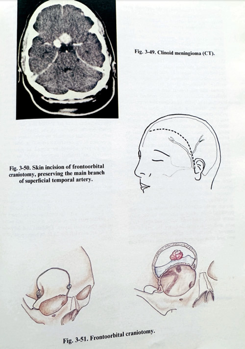
Meningiomas attached on the medial part of the sphenoid (anterior clinoid process), or clinoid meningiomas have been long time considered examples of difficult surgical removal, often associated with complications. Attachment in this area tends easily to involve the internal carotid artery, the optic nerve intracranially and in the optic canal, to invade the neighbouring cavernous sinus and other cranial nerves. The arterial supply of these meningiomas is of multiple sources from the external and internal carotid artery. The important and decisive feature for the radicalism of surgery is the relationship of the tumour to the intracranial carotid artery. Tumour growth from the dura in direct vicinity of where the internal carotid artery enters the subdural space, below and medial to the anterior clinoid, can lead to its encasеment. Here the the arachnoid plays a very important role, if it can be preserved around the carotid, dissection of the arterial wall from the surrounding tumour will be possible. It is the growth of the tumour that does or does not separate the arachnoid of the carotid cistern, and very often the preoperative studies are not able to establish it preoperatively. However, the details of these studies are decisive for the proper orientation during surgery and further on, for its success. These preoperative studies consist of the routine work-up of a meningioma patient: skull X-rays, CT scan of the skull and brain, including enhanced CT, MRI, and angiography. The CT images indicate the location of the tumour mass. They must be complemented with contrast enhanced, thin slice images and bone window CT studies (Fig.3-49). Additional valuable information is obtained from the coronal CT scanning. Coronal slices are better in demonstrating optic canal, intraorbital and intracavernous involvements. MRI gives initial information about the location of internal carotid arteries and the optic nerves, especially if encasement is suspected. MRI is also superior in depicting the peritumoral brain oedema, associated with the lesion. Angiographic features, with selective internal and external contrast injections, reveal the patency of the internal carotid and its branches as well as the tumour's arterial feeders.
As surgery can compromise carotid blood flow, some functional carotid tests, best known when intracavernous presurgical work-up is done, can help us to predict the outcome of temporary or permanent occlusion of the internal carotid during surgery. The first one, the balloon occlusion test, simulates by percutaneous intraarterial technique internal carotid artery occlusion and evaluates its consequences clinically and angiographically. The second is based on the transcranial doppler technique. With the transcranial doppler, the effect of carotid artery compression on the neck can be measured. It should demonstrate collateralisation (reversal of flow in the ipsilateral anterior cerebral artery) and under these circumstances the ipsilateral middle cerebral artery flow can be evaluated. These functional tests have however only a relative predictive value. Of basic importance are also the visual tests; visual affection is the earliest and most important clinical presentation of symptoms in these tumors. Detailed preoperative visual acuity and field studies are essential for preoperative evaluation. Endocrine tests can be a valuable aid for the complete assessment of the patient.
Surgical planning prepares the surgeon for the presumed technical difficulties to be faced during surgery. The craniotomy is usually a standard pterional or frontoorbital one, including adjacent frontal and temporal areas. The epicranial flap for this craniotomy should preserve the superficial temporal artery branches in case they are needed for an STA-MCA microanastomosis (Figs. 3-50; 3-51). The need for some arterial supply interruption has to be decided on in big tumours as detaching the lesion is risky if the attachment is broad, and its proximity to the optic nerve and internal carotid cannot be established visually; however more often this decision must be taken during surgery. Optic foramen and intraorbital involvement will require opening of the optic canal and orbitotomy. However, as details in microanatomy are not easily predictable, any decision must be taken after the lesion has been approached.
The bone flap is either free or attached to the temporal muscle. The flap should include the frontal, temporal and pterional areas, but significant widening of the approach is obtained by orbital wall removal and subperiosteal detachment of the zygomathic arch. We prefer the frontoorbital craniotomy, as the approach gives several advantages: easy direct removal of the lesser wing and control of feeders from the middle meningeal artery extradurally, a wider angle of view, and less retraction of the basal brain surfaces. In all approaches, after lifting the bone flap, follows removal of the lesser and great wing by nibbling and drilling. The removal can continue as much as possible towards the anterior clinoid process.
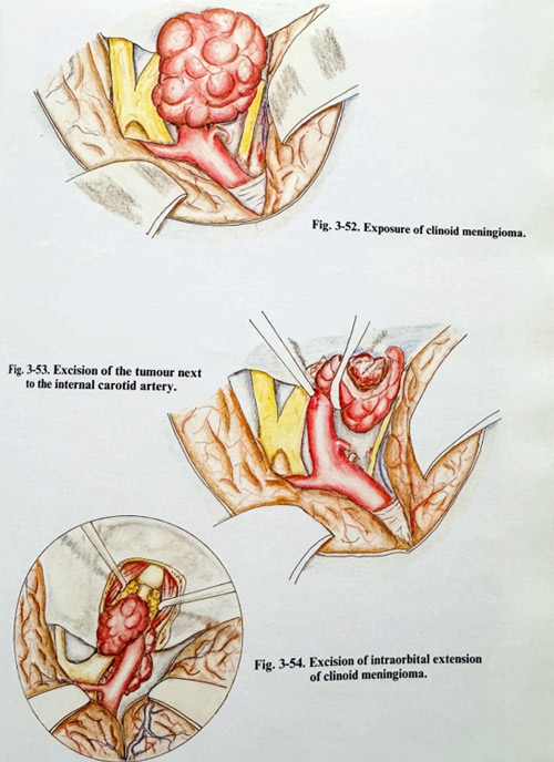
The dura is usually opened along the base after the effect of the applied osmotic agents is obtained. Frontal basal brain surface is retracted, followed by the temporopolar surface. Arachnoid in the Sylvian fissure is opened and the frontal and temporal lobes separated, following the middle cerebral artery branches (Fig. 3-52). In medium and large sized tumours, debulking should be done after this initial exposure. Debulking can be dangerous and it is better to visualise the tumour attachment area well around before cutting or evaporating the tumour mass. Here it is essential to know the exact position of the internal carotid and its major branches to avoid injury during debulking. We are accustomed to alternate dissection in retrograde to the artery's direction with slow debulking (Fig. 3-53). Often distal parts of the carotid, and practically always the middle and anterior cerebral arteries are vested with arachnoid, making the dissection of the arterial wall feasible. If the arterial dissection becomes difficult and risky, the attachment area is carefully inspected for an anterograde approach to the arteries. Optic nerves are usually protected from tumoural invasion by the chiasmatic cistern arachnoid, unless the optic foramen and optic canal dura are the site of the tumour's origin. The optic nerve is dissected first and then the proximal internal carotid can be exposed if this is possible. No attempts should be made to search for major arteries through the tumour, even with well-known artery location, as the invaded adventitia is disrupted easily together with the other arterial layers. If occasional rupture of the wall occurs, the artery should be clipped temporarily on both sides and the defect closed with 8/0 - 10/0 silk suture. If however a major rupture takes place, especially very proximal to the intracavernous portion, the technique of repair is more difficult and time consuming and is similar to that in cases of intracavernous internal carotid sacrifice - venous grafting of the artery or the EC-IC microanastomosis. Which technique to choose is indicated by the results of the balloon occlusion test.
All tumours prone to removal must be excised, the safest approach being with microscissors and bipolar coagu-lation. Affected dura is also excised at the end after being coagulated. Invasions of the orbit or the cavernous sinus are treated in a way appropriate for the location of the residual tumour: with additional exposure by orbitotomy and/or approach to the cavernous sinus (Fig. 3-54). The first surgery should be aimed at maximum removal, as residual tumour mixed with disrupted arachnoid around the major arteries is likely to lead to invasive growth later on. This makes second stage removals practically always incomplete and inefficient, because of this invasion and difficulties in demarcating anatomical landmarks. Once haemostasis is completed, closure is done in the very common way - plastic repairs of dura, rarely of orbital wall, fixation of the bone flap with meticulous readjustment of its orbital part for cosmetic reasons, and suture of the epicranial layers. Epidural and subgaleal drains are also left for 24 to 48 hours postoperatively.