CONVEXITY MENINGIOMAS
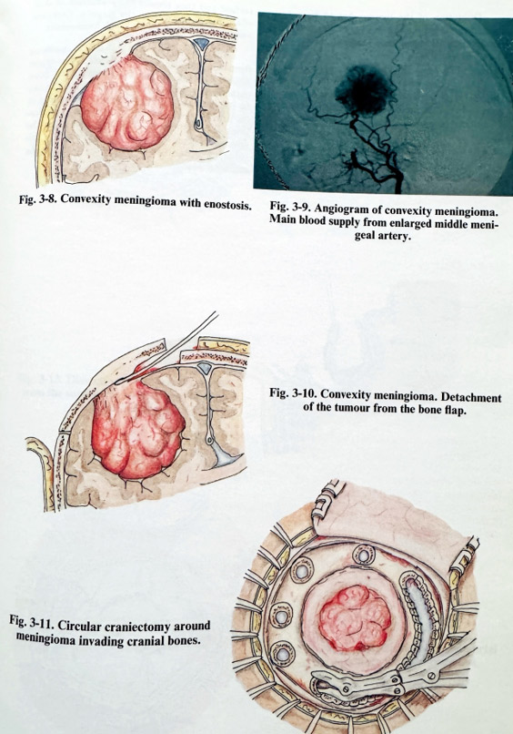
These meningiomas are attached on the dura of the convex surface of cerebral hemispheres and they do not invade any of the dural sinuses. Their shape is spherical or polylobu-lated and they excavate a bed in the cortical surface. They are found more often in the central area, then in the frontal and last in the parietal or temporal areas. At the place of attachment there is often an exostosis or enostosis (Fig. 3-8). Main arterial supply is provided by enlarged meningeal arteries (mainly the middle meningeal artery), but epicranial arteries can contribute in the case of exostoses or skull bone infiltration. Cortical arteries can also give off branches to the tumour (Fig. 3-9).
The craniotomy is planned to expose the tumour attachment area in the centre of the exposed dura. An exostosis or infiltration should be included in the preplanned flap. Bone cutting lines should be within normal bone, outside the tumour involvement. Sufficient space for manipulation of 1-2 cm should be provided between the attachment area and the bone edge.
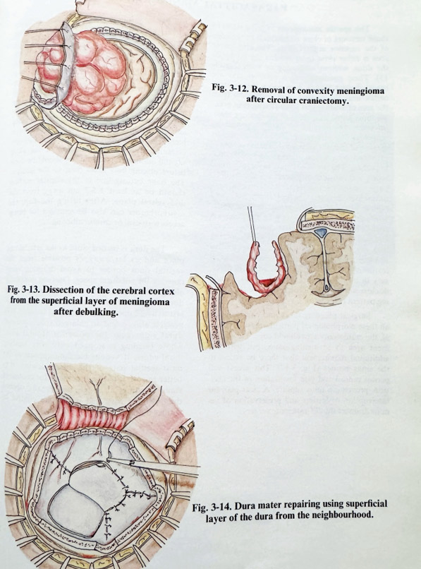
Craniotomy is performed in the standard way, including special precautions to avoid excessive bleeding, and there can be more than the usual number of burr holes. The flap is lifted after careful separation of the infiltrated dura and tumour from the inner flap surface. A fine periosteal elevator or dissector is used to scrape the inner table to avoid pulling of the tumour (and in so doing, also the important adjacent neural and vascular structures) (Fig. 3-10). Haemostasis after the flap lift is the most important step of the craniotomy; it requires skilful handling by the surgeon and should prevent excessive bloodloss at this early stage of the operation. Bleeding dural arteries are coagulated with bipolar coagulation, vascular bony channels are waxed and bleeding under the craniotomy edges is packed with oxidised cellulose. All abnormal bone must be removed. In some cases of extensive meningioma infiltration and growth inside the cranial bone, lifting a flap is rather risky, as severe haemorrhage and brain injury may occur. These tumours are approached with a circular craniectomy done with the high speed drill or with multiple burr holes and nibbling, interrupted periodically for haemostasis by waxing. After the lesion is encircled, it can be dissected at the dural level. If epidural dissection is inefficient, the dura is opened in a circular way close to the affected bone, the procedure followed being that for routine removal (sometimes with all the tumour "en block") (Figs. 3-11; 3-12).
The dura is opened in the standard way for a meningioma: first at a point which is non-adherent to the underlying cortex area, and then as close as possible to the attachment. Dural edges are held on traction sutures. If an enlargement is needed for safe manipulation around the tumour, short radial additional cuts can be added.
Debulking is started through the attachment place after biopsy sample taking. Marginal parts of the tumour are preferred for biopsy. In debulking a convexity meningioma additional bleeding appears from cortical branches to the tumour when penetrating into the depth of the tumour. They are carefully stopped with bipolar coagulation, especially when over a thin remaining layer of tumour. Debulking continues until the remaining tumour layer becomes flexible.
Dissection of the tumour has to be performed under magnification and, as a rule, with preservation of arachnoid. Manipulation is at the sacrifice of the tumour, protecting the cortical surface. The tumour is retracted more easily with the traction sutures of the dura on its side (Fig. 3-13). If the subarachnoid space is opened, arterial branches are followed until they enter the tumour before being coagulated and divided.
Special attention, as in parasagittal tumours, is paid to cortical venous drainers, whose damage is often the cause of postoperative focal deficit.
Some tumours are more adherent to the cortical surface and they require the maximum of skill, effort and patience from the surgeon. Complete removal is followed by thorough haemostasis.
There is always a dural defect resulting from this removal, and it is closed in one of the accepted methods (Fig. 3-14). Infiltrated bone tissue is removed. A normal part of the flap can be retained and the remaining defect closed at the same surgical session if there are no indications for significant brain oedema after the removal (steroids have to be considered for this surgery). All other layers are closed in the common way.