PARASAGITTAL MENINGIOMAS
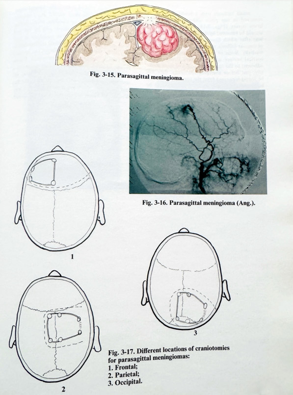
This specific meningioma location refers to those tumours in close relationship to the edge of the superior sagittal sinus. The attachment area is either close to or reaches the border of the sinus, without infiltrating its walls (Fig. 3-15). These commonly seen meningiomas are round-shaped lesions excavating the cerebral cortex near the midline, and originating from the area of arachnoidal villae (Pachionian granulations) found in vicinity of the superior sagittal sinus. The tumour is mainly supplied by pathologically enlarged branches of the middle meningeal artery. Additional arterial supply is provided by cortical, falcine and epicranial branches. The lesions often involve cranial bones and can seriously affect the venous drainage, changing the pattern of venous flow through parasagittal, cortical anastomotic and diploic veins. Not infrequently, without really reaching the midline, the area of attachment is next to broad venous lacunae collecting cortical and tumour venous drainers and widely opening into the superior sagittal sinus. Preoperative imaging should reveal all these morphological relationships and angiography, which is necessary in all, except very small tumours, should disclose not only the feeders, but also the draining pattern (Fig. 3-16).
Surgical planning takes into consideration all these morphological particularities. In general, the craniotomy area should cover the attachment area as in meningiomas, but with the additional requirement that there be access to the sinus border (Fig. 3-17). This access will permit radical and safe finalization of the excision, preventing sinus rupture. A clear plan for interruption of feeders and preservation of venous drainers should anticipate surgery.
Patient positioning can be supine, oblique, lateral, prone, or semi-sitting, depending on the location of the lesion. The routine measures to avoid venous congestion are essential in this location of the meningioma. Epicranial incisions are designed in the common way, although they can be extended to the contralateral side for functional or cosmetic reasons. Bone flaps are performed in the manner used in convexity meningiomas with particular attention to the medial border of the craniotomy. In this place any dural injury can provoke massive venous bleeding. Therefore, some technical tips are essential. Certain identification of the skull sagittal midline (sagittal suture) should be done before placing burr holes close to the midline.
The bone cutting line to the sagittal midline should be at least 1.5-2 cm away from the midsagittal plane. After lifting the flap, the sagittal border can also be removed for better access with drill or careful nibbling.
The dura is incised close to the attachment place and as far away as possible from the sagittal sinus border to avoid draining veins crossing the subdural space. Opening continues along the attachment border, sparing intact dura. The most medial part of opening is left last to avoid opening venous dural lacunae. It is possible also to find an enlarged sphenoparietal dural sinus near the posterior frontal area, whose bleeding is stopped only after bending dural edges. If venous bleeding appears at the most medial part of the dural incision, it is better to stop it with all methods for control of sinus bleeding, although the already opened dura permits easy and immediate control by sutures or clips.
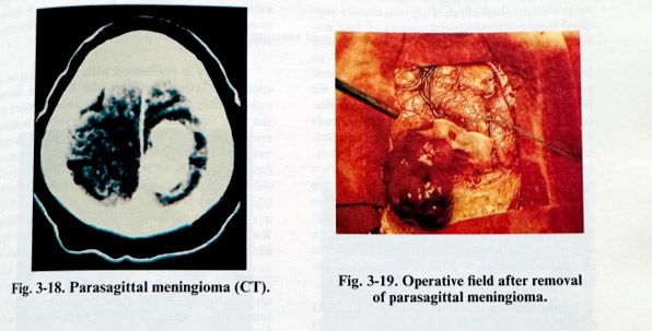
Small tumours with a broad base can be removed in one piece without damaging the brain cortex (Figs. 3-18; 3-19). Big tumours should be removed by debulking in the common way after which dissection of the residual layer of tumour follows, starting laterally and moving medially. Some of the venous collectors on the cortical surface are found very stretched and adherent to the tumour. If followed most medially, just before emerging epidurally, they will be found separated from the tumour. There can be found the proper plane of dissection to be followed. If the laser has to be the tool of removal, veins are cautiously and completely covered with soaked cottonoids. Special attention is paid to the most medial dural involvement, where, because of bleeding, small residual areas infiltrated with tumour can be left and cause early recurrence. It is necessary to perform at least the steps of residual tumour dissection under the surgical microscope, i.e., revision for radical removal, and haemostasis. Plastic repair of dura follows, aimed at attaining a watertight closure. After all affected bony tissues have been removed, the closure of the flap and /or plastic repair are done in the usual way. Wound is drained and the epicranial flap closed.